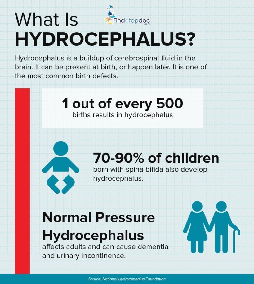
Hydrocephalus is a condition that occurs when fluid builds up in the skull and causes the brain to swell. The name means “water on the brain.” Brain damage can occur as a result of the fluid buildup. This can lead to developmental, physical, and intellectual impairments. It requires treatment to prevent serious complications. Hydrocephalus mainly occurs in children and adults over 60, but younger adults can get it too. The National Institute of Neurological Disorders and Stroke (NINDS) estimates that 1 to 2 of every 1,000 babies are born with hydrocephalus.
Causes
Cerebrospinal fluid (CSF) flows through your brain and spinal cord in normal conditions. Under certainconditions, the amount of CSF in your brain increases. The amount of CSF can increase when:
- a blockage develops that prevents CSF from flowing normally
- there is a decrease in the ability of blood vessels to absorb it
- your brain produces an excess amount of it
Too much of this fluid puts your brain under too much pressure. This pressure can cause brain swelling, which can damage your brain tissue.
When hydrocephalus occurs in adults, CSF levels rise but the amount of pressure is usually normal. It still causes the brain to swell and can lead to impaired functioning. In adults, this condition usually results from conditions that prevent CSF from flowing. However, in some cases, there is no known cause.
You might be at higher risk if you have experienced any of the following:
- brain-related infections such as meningitis
- head injuries
- bleeding from a blood vessel in your brain
- brain surgery
Diagnosis
If you suspect that you or your child has hydrocephalus, your doctor will perform a physical exam to look for signs and symptoms. In children, doctors check for eyes that are sunken in, slow reflexes, a bulging fontanel, and a head circumference that is larger than normal for their age. Your doctor may also use an ultrasound to get a closer look at the brain. These tests use high-frequency sound waves to create images of the brain. Magnetic resonance imaging (MRI) scans can be used to look for signs of excess CSF. MRIs use a magnetic field and radio waves to make a cross-sectional image of the brain. Computerized tomography (CT) scans can also help diagnose hydrocephalus in children and adults. CT scans use several different X-rays to form a cross-sectional image of the brain. These scans can show enlarged brain ventricles that result from too much CSF.
Treatment
Hydrocephalus can be fatal if it’s left untreated. Treatment may not reverse brain damage that’s already occurred. The goal is to prevent further brain damage. This involves restoring the normal flow of CSF. Your doctor may explore either of the following surgical options:
Shunt Insertion
In most cases, a shunt is surgically inserted. The shunt is a drainage system made of a long tube with a valve. The valve helps CSF flow at a normal rate and in the right direction. Your doctor inserts one end of the tube in your brain and the other end into your chest or abdominal cavity. Excess fluid then drains from the brain and out the other end of the tube, where it can be more easily absorbed. A shunt implant is typically permanent and has to be monitored regularly.
Ventriculostomy
A procedure called a ventriculostomy can be performed as an alternative to having a shunt inserted. This involves making a hole at the bottom of a ventricle or in between ventricles. This allows CSF to leave the brain.
The outlook for someone suffering from hydrocephalus depends largely on the extent of the symptoms. Many children suffer lifelong brain damage. By working with professionals including pediatricians, special education teachers, mental health providers, occupational therapists, developmental therapists, and pediatric neurologists, children can learn to manage their disability and lessen the lifelong effects.